


Background
50 year old male with incidental detection of mass in right kidney on ultrasound. Partial nephrectomy done thereafter.
Gross specimen
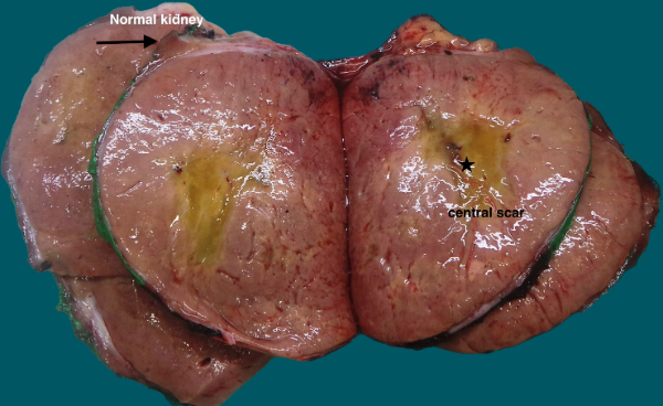
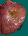
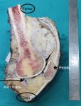
Fig. 17a; Gross: Well circumscribed tumor with mahogany brown cut surface and central scar
Microscopy
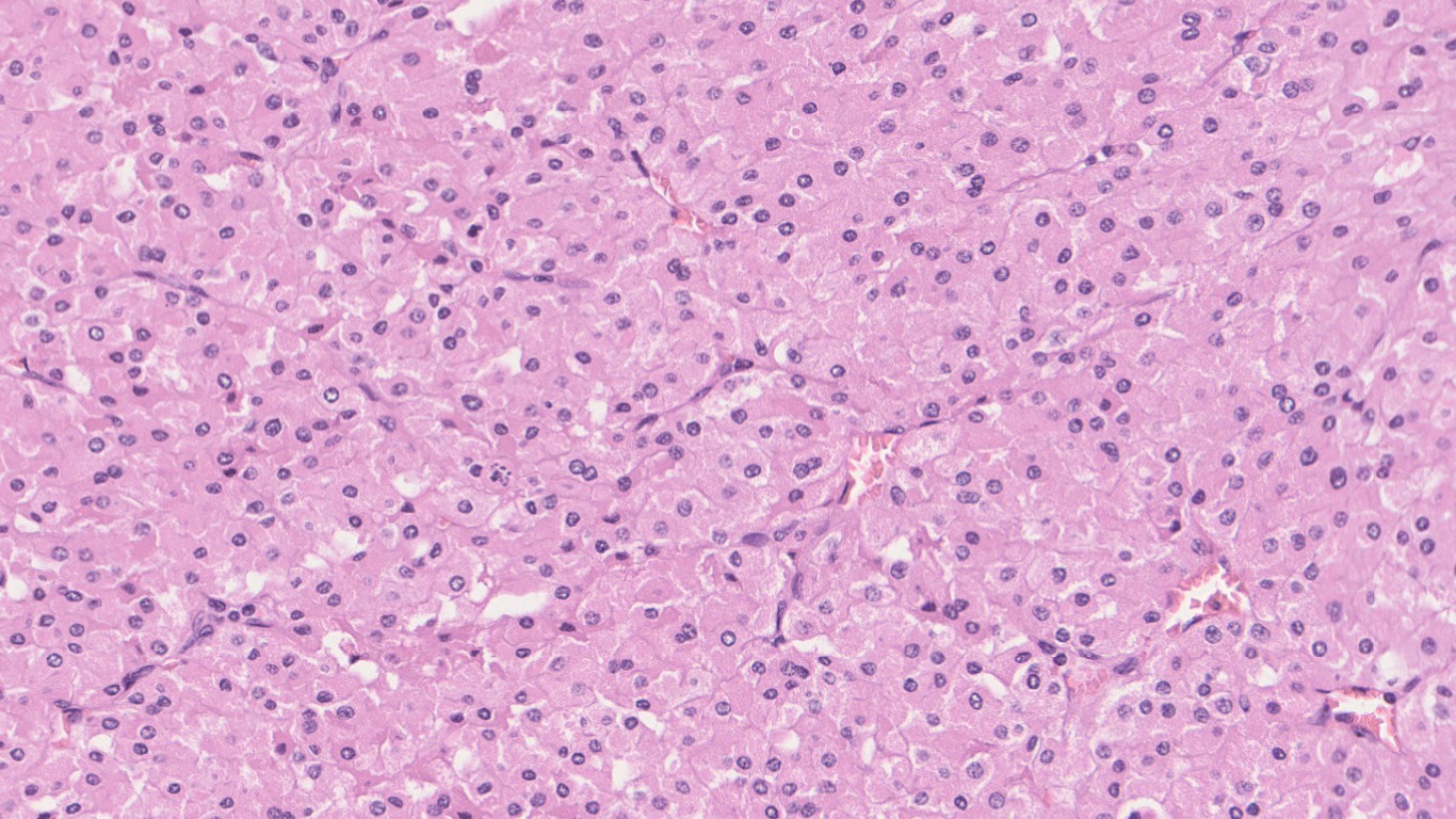
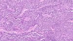
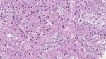
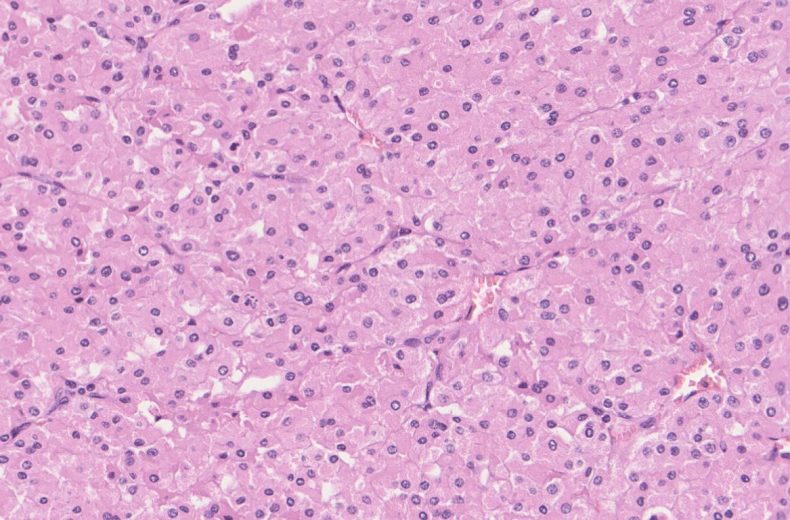
Fig 17b; H&E; 20x

Fig 17c; H&E; 20x
Immunohistochemistry
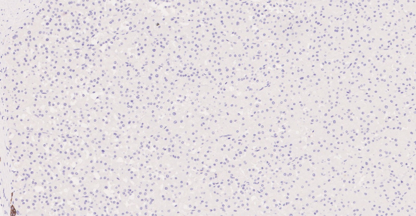
Fig 17d; CK7

Fig 17e; AMACR

Fig 17f; CD117

Fig 17g; SDHB

Fig 17h; E-Cadherin
Questions & Answers:
- What is your diagnosis?
- Oncocytoma
- Chromophobe Renal Cell Carcinoma
- Succinate Dehydrogenase deficient RCC
- Low grade Oncocytic tumor
- Which of the following is true for above tumor?
- Strong CK7 and AMACR positivity
- Subset of cases are CD117 negative
- Positive for Hale colloidal iron staining
- Negative for Low molecular weight cytokeratin
- Hybrid case of Oncocytic tumor and Chromophobe RCC is seen in:
- Birt-Hogg-Dube Syndrome
- Tuberous Sclerosis
- VHL syndrome
- MIT Family translocation RCC
- Oncocytoma is a benign epithelial tumor with solid, solid-nested, or cystic architecture
- Incidence: Constitute 5-9% of all renal cell neoplasm
- Age: Peak at 7th decade
- Clinical findings: Most of the cases are asymptomatic and are detected incidentally on radiological investigations for some other cause
- Location:
- Predominantly cortex based
- Large tumors can expand into medulla, renal sinus and even large renal vein branches
- Gross features:
- Cut surface: Mahogany brown to tan or yellow with central scar
- Microscopic features:
- Solid- nested architecture is characteristic
- Rarely small papillary protrusion into renal tubules or in cystic areas
- Composition of microcyst and macrocyst can occur
- Some cases can show degenerative changes in the form of bizarre cells with pleomorphic nuclei
- Immunohistochemistry:
- Positive stains: CD117, E-cadherin, Pancytokeratin & Low-molecular weight cytokeratin
- Rarely positive for: CD10 & AMACR
- Negative stains: CK7, Vimentin (may be positive in small cells around the central scar)
- Hale colloidal iron staining is negative but can show positivity in luminal aspect of cells
- Genetic susceptibility:
- Sometimes can be seen in association with Birt-Hogg-Dube Syndrome: Have hybrid Oncocytic and Chromophobe tumor
- Differential Diagnosis:
- Chromophobe Renal cell carcinoma: Positive for CD117 & CK7, while are negative for Vimentin
- Low Grade Oncocytic tumor: Positive for CK7, while are negative for CD117
- Succinate dehydrogenase deficient RCC: Negative for CD117 & CK7. SDHB expression is lost
Contributed by: Dr. Meenakshi Kamboj
Compiled by: Dr. Diksha Karki
In case of queries, email us at: info.pathbliss@rgcirc.org
Kidney Neoplasms Oncocytoma Renal tumors
Last modified: 08/09/2021