


Background
35-year- old male, presented with left second toe swelling. MRI showed a solid soft tissue mass measuring 4.1 x 2.8cm, showing moderate homogenous enhancement. Trucut biopsy sent for histopathological examination.
Microscopy
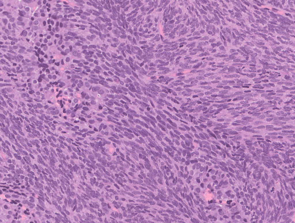
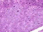
Fig.1a; H&E; 20x
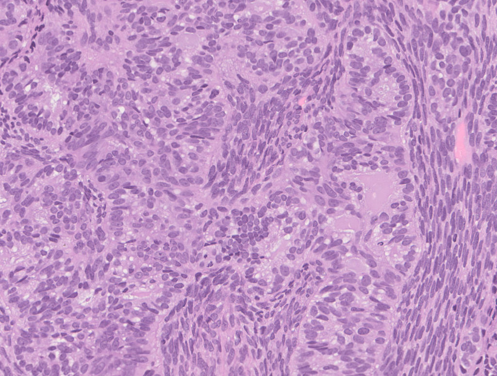
Fig.1b; H&E; 20x
Biopsy showed a high grade tumor with biphasic pattern. One component was of a high-grade sarcoma [Fig.1a] and intimately admixed were epithelial components [Fig.1b] in the form of glands and solid nests.
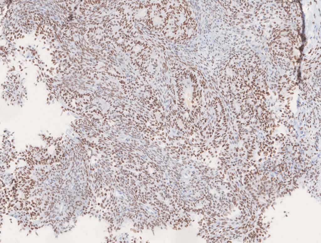
Fig.1c; TLE-1
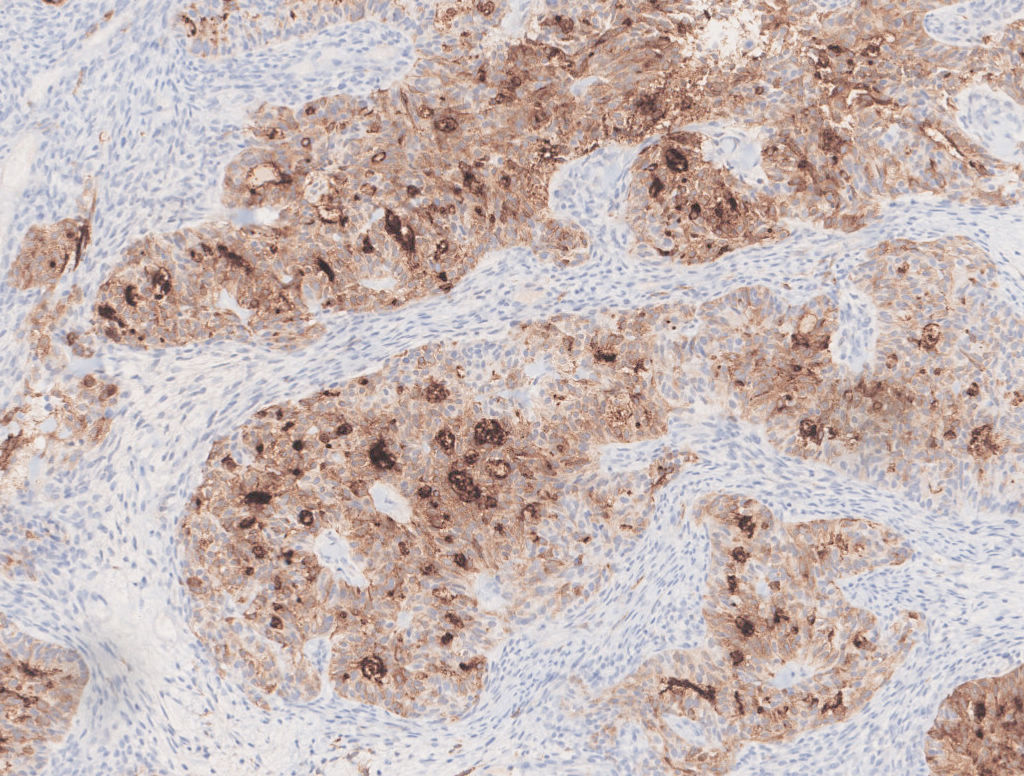
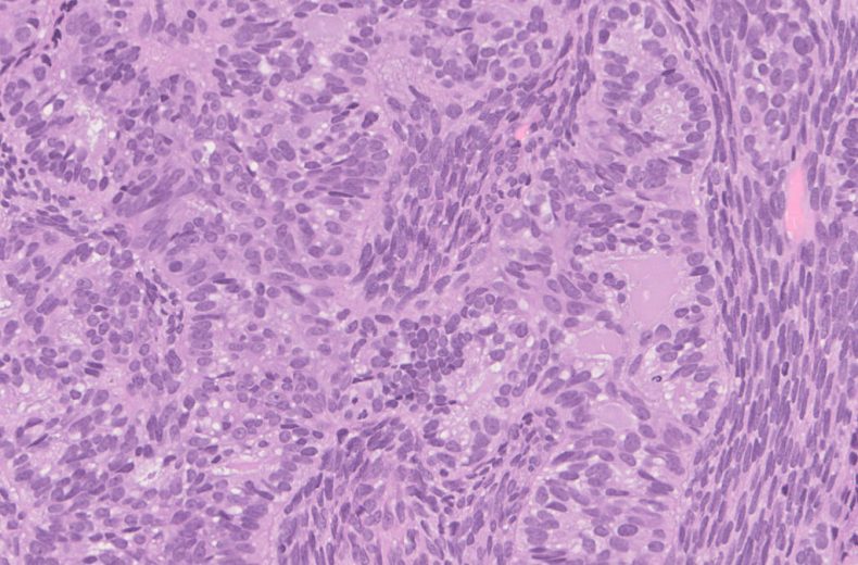
Fig.1d; EMA
Immunohistochemistry revealed TLE, EMA & CK positivity [Fig.1c&d].
Final Impression: Biphasic Synovial Sarcoma.
Contributed by: Dr. Anila Sharma
Compiled by: Dr. Ankur Kumar & Dr. Himanshi Diwan
In case of queries, email us at: kumar.ankur@rgcirc.org
Soft tissue tumors Synovial sarcoma
Last modified: 05/06/2021