


Background
54-year-old male, with history of scleroderma on methotrexate, presented with right frontal scalp ulcerated lesion measuring 3 x 1 cm in size. Punch biopsy from lesion.
Microscopy

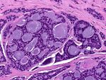
Fig.2a; H&E 2.5x

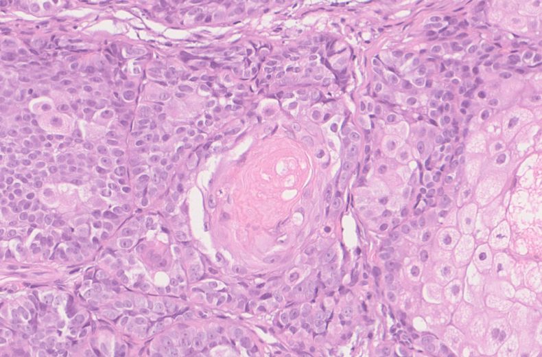
Fig.2b; H&E; 20x

Fig.2c; H&E; 20x
Images show ulcerated epidermis with a dermal tumor. The tumor has a lobulated appearance with pushing margins [Fig.2a]. The tumor cells are polygonal with bubbly or eosinophilic cytoplasm having oval vesicular nuclei exhibiting conspicuous nucleoli and brisk mitosis [Fig.2b]. Squamous metaplasia with keratin pearl formation is also seen [Fig.2c].
Final Impression: Sebaceous Carcinoma, Grade 1
- Origin: Sebaceous glands of skin or ocular adnexa
- Types: Periocular or Extraocular
- Periocular tumors: more aggressive
- May follow radiation therapy
- Association with Muir Torre syndrome
- Necrosis is related to poor prognosis
- IHC:
- Positive stains: CK, EMA, LeuM1, BEREP4, Androgen Receptor
- Negative stains: CEA, s100
- Differential diagnosis:
- Basal cell carcinoma with sebaceous differentiation
- Squamous cell carcinoma with hydropic changes
Contributed by: Dr. Anila Sharma
Compiled by: Dr. Ankur Kumar & Dr. Himanshi Diwan
In case of queries, email us at: kumar.ankur@rgcirc.org
Head & Neck Sebaceous carcinoma Skin adnexal tumor
Last modified: 05/06/2021