Background
A 56-year-old female presented with postmenopausal bleeding. MRI pelvis showed bulky uterus with large myometrial fibroid having cystic degeneration. Total abdominal hysterectomy was done. Sections from the myometrial mass.
Microscopy
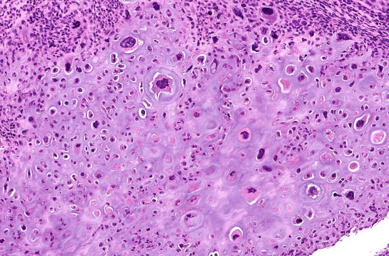
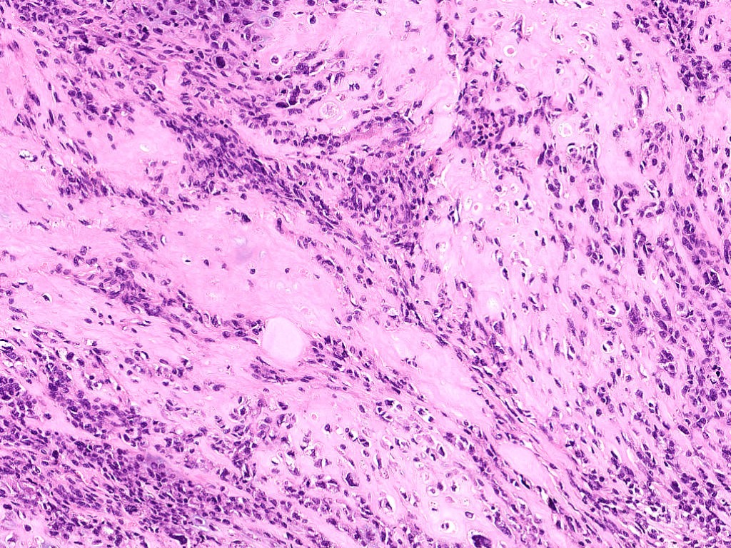
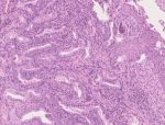
Images show areas of high-grade spindle cell sarcoma in fascicles and sheets [Fig.12a] with focal areas of myogenic differentiation [Fig.12b]. Many multinucleated tumor cells are seen [Fig.12c] along with sheets of epithelioid cells in myxoid background [Fig.12d]. Areas of heterologous differentiation in the form of osteosarcoma [Fig.12e] and chondroid differentiation [Fig.12f] are also noted.
No epithelial component is readily identified.
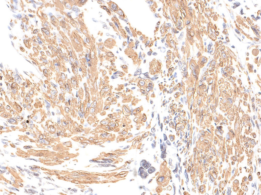
On Immunohistochemistry, tumor cells are diffusely negative for CK [Fig.12g]. Areas with myogenic differentiation show positivity for SMA [Fig.12h] and Desmin [Fig.12i].
Final Impression: High Grade Leiomyosarcoma With Heterologus (Osteosarcoma/Chondrosarcoma) Differentiation.
- Uterine leiomyosarcoma is a malignant mesenchymal tumor derived from smooth muscle cells
- Age of presentation: 4th-5th decade of life
- Location: Uterine corpus (common), cervix
- Germline mutation in Fumarate Hydratase⇒ increased risk of developing uterine leiomyosarcoma
- Genetic alteration: Frequently mutated genes:
- TP53
- ATRX
- MED12
- Subtypes:
Spindle cell leiomyosarcoma |
Epithelioid leiomyosarcoma |
Myxoid leiomyosarcoma |
|
|
|
- Essential diagnostic criteria:
Spindle cell leiomyosarcoma(Two or more of the following) |
Epithelioid leiomyosarcoma(One or more of the following) |
Myxoid leiomyosarcoma(One or more of the following) |
|
|
|
- Rare cases with osseous and chondro-osseous differentiation have been reported in literature
- Immunohistochemistry (Positive stains):
- Desmin, h-caldesmon, SMA, CD10
- EMA, CK (epithelioid leiomyosarcoma)
- ER, PR, p16, p53 overexpression (Spindle cell leiomyosarcoma)
- Differential diagnosis:
- Smooth muscle tumor of uncertain malignant potential (Criteria):
- Tumor cell necrosis: absent, focal moderate atypia, ≥5 mitosis/10HPF or,
- Tumor cell necrosis: absent, diffuse severe to moderate atypia, 5-9 mitosis/10HPF or,
- Tumor cell necrosis: present, none to mild atypia, <10 mitosis/10HPF
- Atypical leiomyoma (Criteria):
- Tumor cell necrosis- Absent
- Focal moderate to severe atypia
- <5mitosis/10HPF
- Mitotically active leiomyoma (Criteria):
- Tumor cell necrosis- Absent
- None to mild atypia
- ≥5mitosis/10HPF
- Undifferentiated endometrial sarcoma:
- Endometrium and myometrium location
- Negative for Desmin, caldesmon and Actin
- Rhabdomyosarcoma:
- Rare in uterus
- Search for rhabdomyoblast.
- Positive for Desmin and Myogenin
- Negative for caldesmon
- Smooth muscle tumor of uncertain malignant potential (Criteria):
- Prognosis:
- Aggressive, even with tumor confined to uterus
- Recurrence (50-70%) occur in lung and in pelvis
- Progression depends on stage
- Size of neoplasm < 5cm (in Stage I tumor) and premenopausal women have more favourable outcome as seen in some studies
- Treatment:
- Postmenopausal women: Early-stage: TAH and BSO
- Removal of ovary and lymph node is controversial, as metastasis to these organs occur in small percentage of cases
- Premenopausal women: therapy is controversial
- In stage I and II cases, role of adjuvant systemic therapy or radiotherapy on survival is uncertain
- Some patients respond to hormonal therapy
Contributed by: Dr Sunil Pasricha
Compiled by: Dr Diksha Karki
In case of queries, email us at: kumar.ankur@rgcirc.org
Female genital tract Heterologous differentiation Leiomyosarcoma Smooth muscle tumor Soft tissue tumors
Last modified: 28/06/2021
