


Background
1-year-old male, presented with soft tissue mass in left pre-auricular region, since 5 months. Radiology showed a 3.6 x 2.8cm, soft tissue mass, in the left temporo-parietal region extending into maxilla and orbit. Biopsy was sent for histopathological examination.
Microscopy

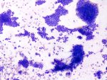
Fig.4a; H&E; 0.75x

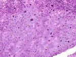
Fig.4b; H&E; 10x

Fig.4c; H&E; 20x

Fig.4d; H&E; 20x

Fig.4e; H&E; 20x
Images show tissue cores infiltrated by a poorly differentiated tumor [Fig.4a & b]. Tumor cells are arranged in diffuse sheets and have large hyperchromatic nuclei, coarse chromatin, inconspicuous nucleoli and scant cytoplasm, exhibiting brisk mitosis [Fig.4c & d]. Some areas show sheets of these neoplastic cells exhibiting rhabdoid morphology [Fig.4e].

Fig.4f; LCA

Fig.4g; NKX2.2

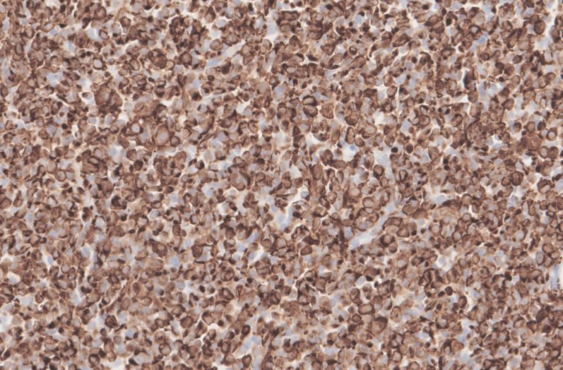
Fig.4h; DESMIN

Fig.4i; MYOGENIN
Immunohistochemistry showed no expression with LCA [Fig.4f] & NKX2.2 [Fig.4g], while strong expression was noted with Desmin [Fig.4h] & Myogenin [Fig.4i].
Final Impression: Embryonal Rhabdomyosarcoma.
- Age group: Children and young adults (3-12 years)
- Sites:
- Head and neck (particularly orbit, nasopharynx, middle ear, oral cavity)
- Retroperitoneum, Genitourinary tract, Bile ducts.
- Most common sites for metastasis:
- Bone marrow, lungs, soft tissue, serosal surface
- Variants:
- Sarcoma botyroides
- Embryonal Rhabdomyosarcoma with anaplastic features
- Immunohistochemistry:
- Positive stains: Myogenin (most sensitive and specific), MyoD1 & Desmin
- CK and Neuroendocrine markers can be positive.
- Molecular:
- Complex karyotypes(+2,+8,+13)
- Loss of 11p15
- Prognosis:
- Anaplasia is a negative prognostic factor, regardless of whether it is focal or diffuse.
- Differential diagnosis:
- Alveolar and pleomorphic rhabdomyosarcomas
- Desmoplastic small round cell tumor
- Ewing/ PNET
- Lymphoma
- Monophasic synovial sarcoma
- Neuroblastoma
- Wilms tumor
Contributed by: Dr. Meenakshi Kamboj
Compiled by: Dr. Ankur Kumar & Dr. Himanshi Diwan
In case of queries, email us at: kumar.ankur@rgcirc.org
Embryonal rhabdomyosarcoma Head & Neck Pediatric tumors Rhabdomyosarcoma Soft tissue tumors
Last modified: 05/06/2021