


Background
31-year-old male, presented with left thumb swelling. Swelling was circumscribed nodular and red in color measuring 1.7×1.5cm. Radiology was suggestive of Giant cell tumor of Tendon sheath. Wide local excision was done and tissue was sent for histopathological examination.
Microscopy
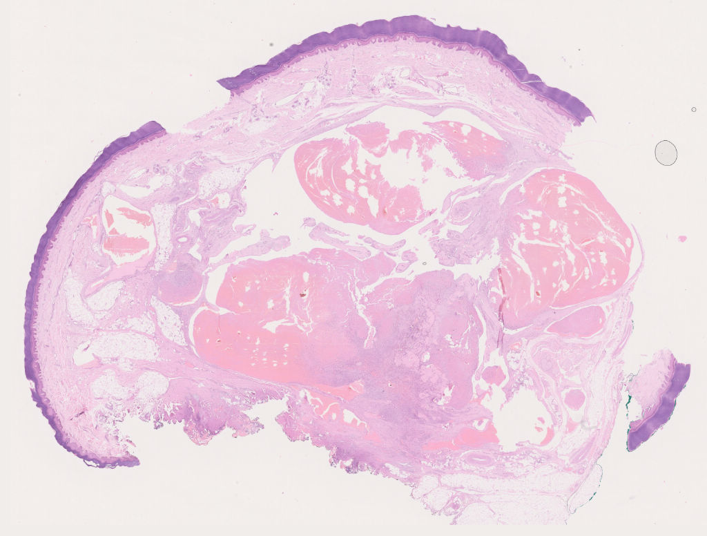
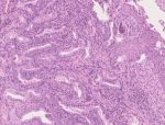
Fig.3a;H&E; 0.42x
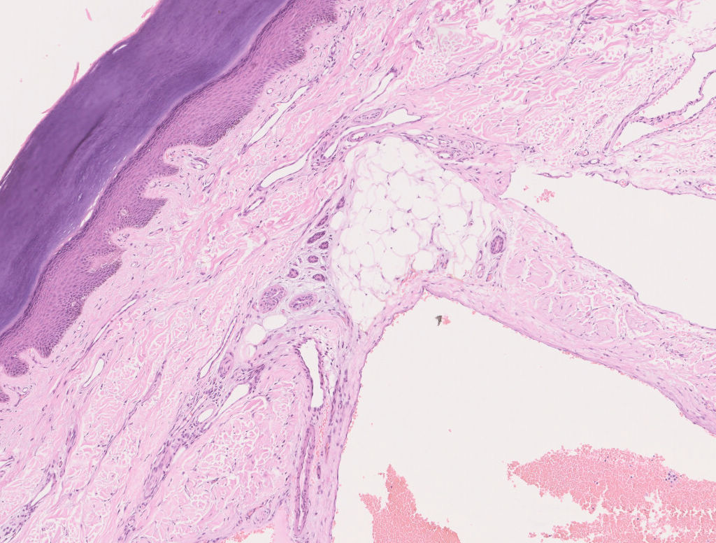
Fig.3b; H&E; 5x
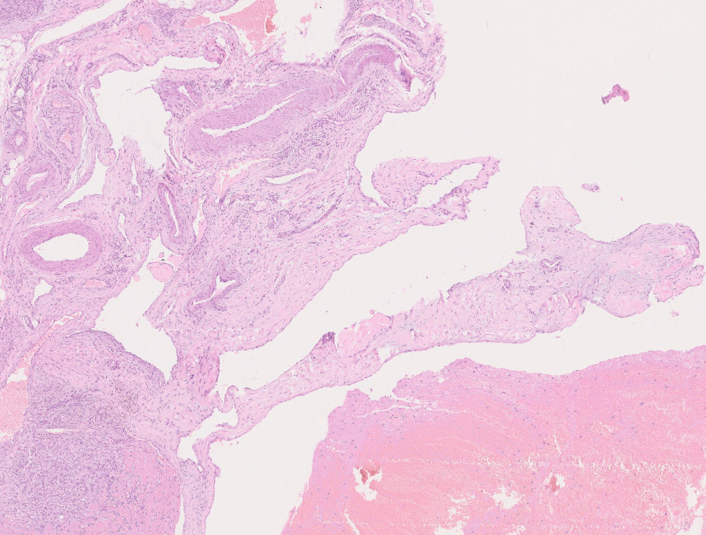
Fig.3c; H&E; 2.5x

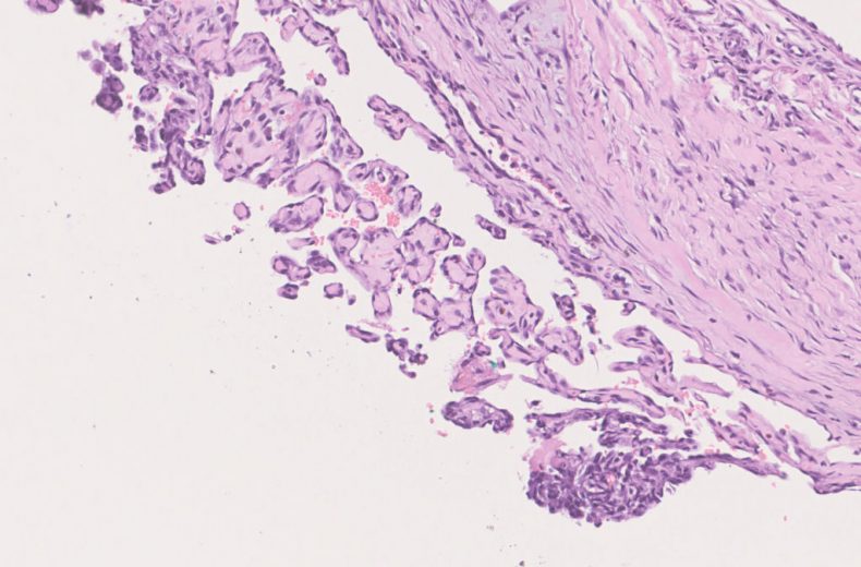
Fig.3d; H&E; 10x
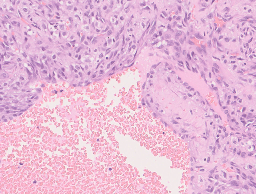
Fig.3e; H&E; 20x
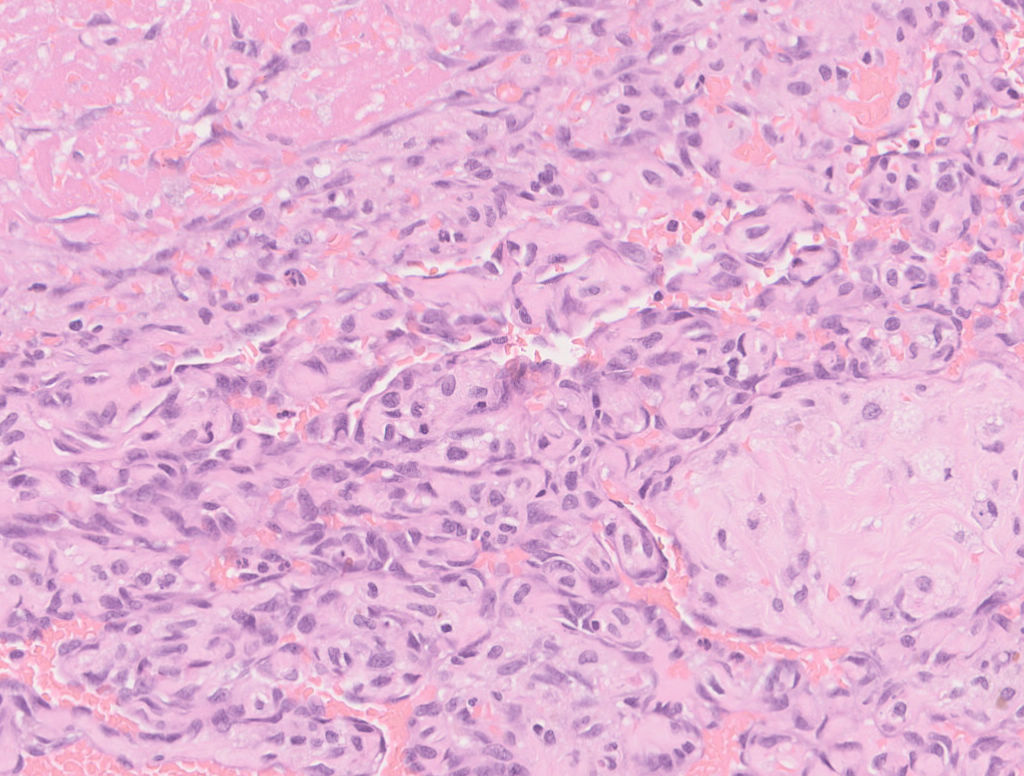
Fig.3f; H&E; 20x
Images show hyperkeratotic and acanthotic stratified squamous epithelium with underlying dermis and subcutaneous tissue showing large blood filled spaces [Fig.3a-c]. On higher magnification an intravascular tumor with anastomosing vascular channels filled with blood and lined by spindle shaped cells with spindle shaped nuclei is noted with focal pseudopapillary pattern. [Fig.3d-e]. Thrombus and fibrin deposition also seen [Fig.3f].
Final Impression: Papillary Endothelial Hyperplasia.
- Reactive condition
- Also known as Masson’s tumor
- Represents exuberant organization and recanalization of thrombus
- Common Sites: Dermis and subcutis of head and neck, fingers & trunk
- Immunohistochemistry:
- Positive stains: SMA, CD34, FactorVIII
- Commonly confused with Angiosarcoma.
- Intravascular location, absence of frank necrosis or marked pleomorphism or mitosis negates Angiosarcoma.
Contributed by: Dr. Sunil Pasricha
Compiled by: Dr. Ankur Kumar & Dr. Himanshi Diwan
In case of queries, email us at: kumar.ankur@rgcirc.org
Non neoplastic Papillary endothelial hyperplasia Soft tissue lesion
Last modified: 05/06/2021