


Background
A 77-year-old lady presented with postmenopausal bleeding since 3 months. MRI showed a heterogenously enhancing lesion measuring 1×0.9cm, involving external os. Biopsy from the lesion was sent for histopathological examination.
Microscopy
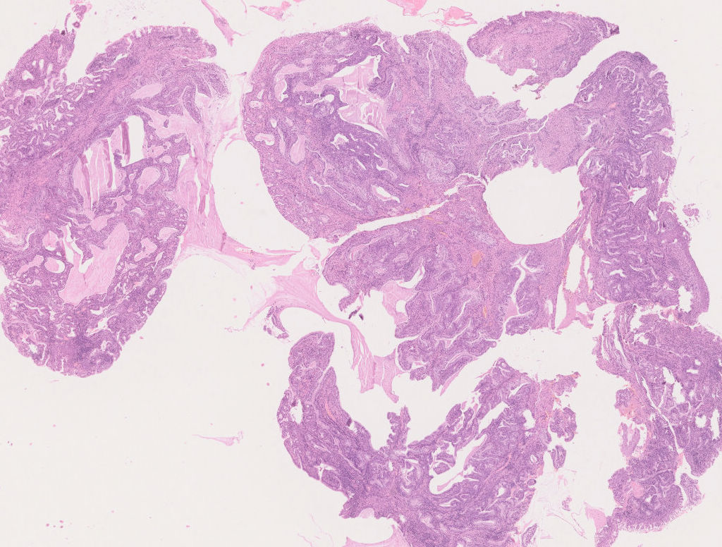
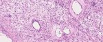
Fig.6a; H&E; 1.25x
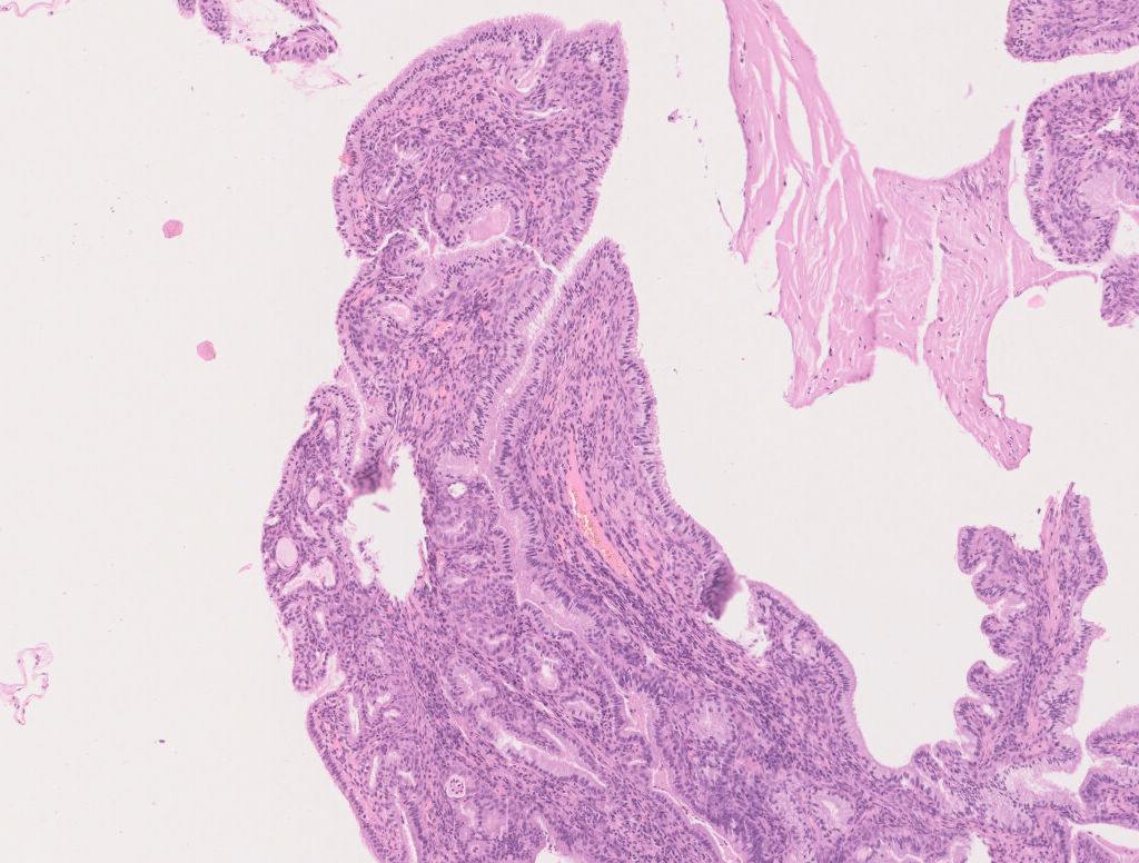
Fig.6b; H&E; 5x
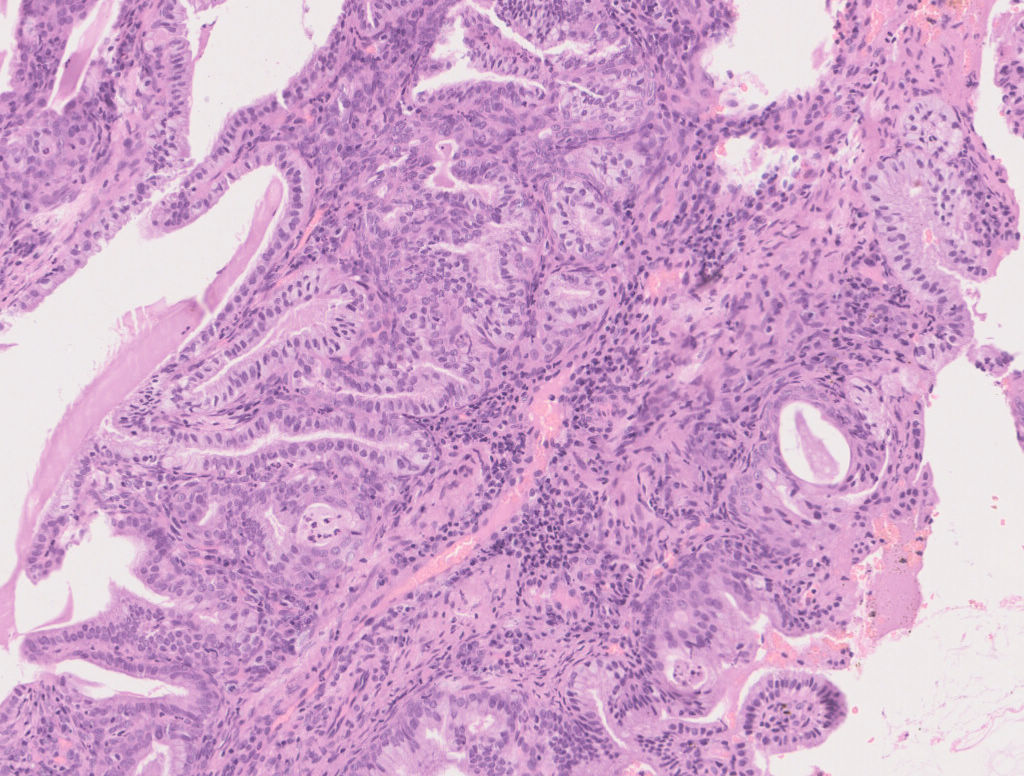
Fig.6c; H&E; 10x
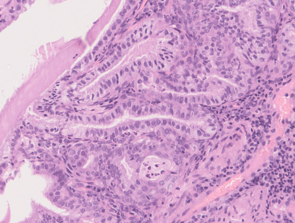
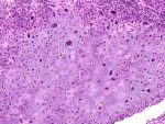
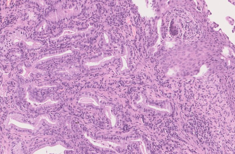
Fig.6d; H&E; 20x
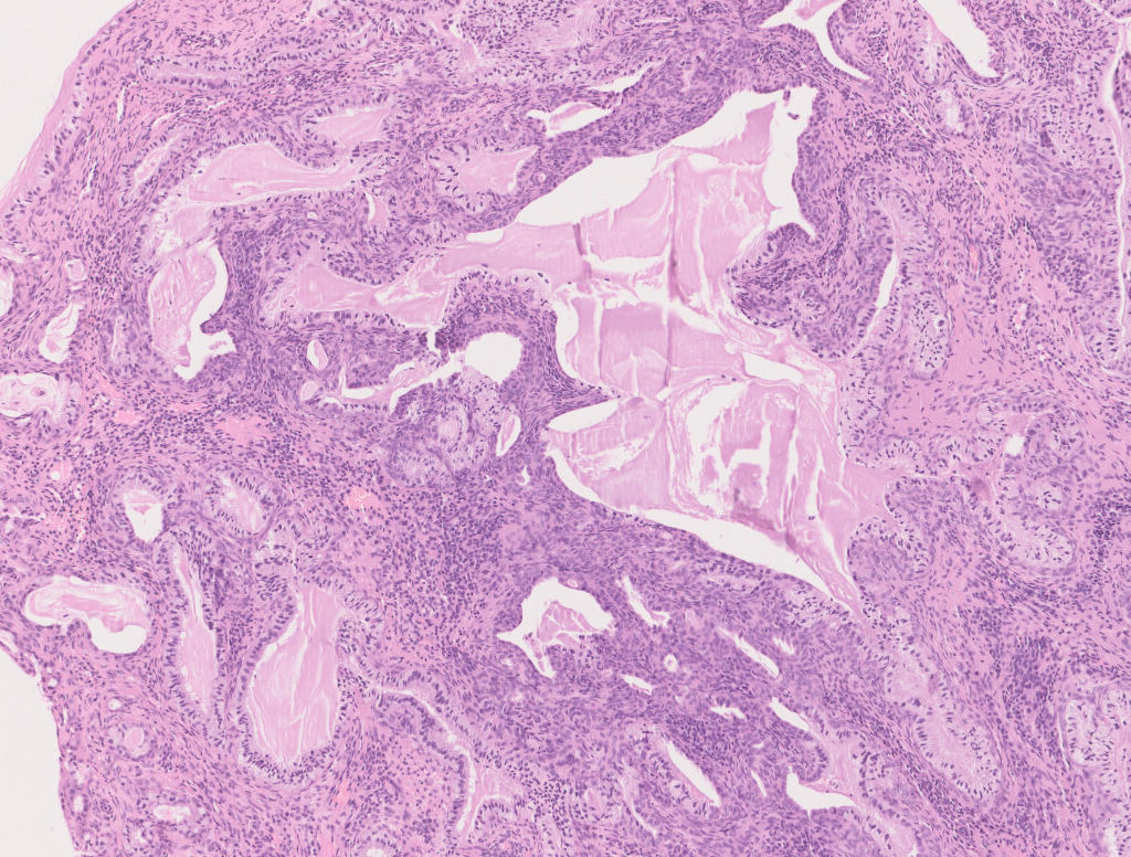
Fig.6e; H&E; 5x
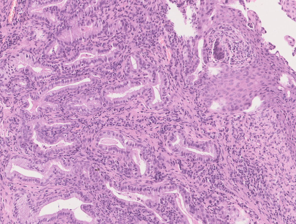
Fig.6f; H&E; 10x
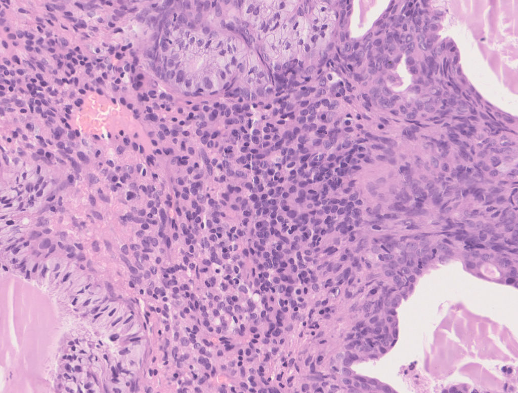
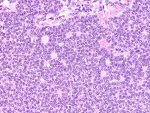
Fig.6g; H&E; 20x
Images show polypoidal tissue cores lined by endocervical mucosa with deep invaginating proliferating glands, lined by tall columnar mucinous epithelium [Fig.6a-d]. Cystic structures are seen formed by pseudoglandular lumina, filled with secretions [Fig.6e]. Focal scattered areas of squamous metaplasia seen on surface [Fig.6f]. Intervening stroma is cellular [Fig.6g]. There was no evidence of nuclear atypia or malignancy. No apical mitosis identified.
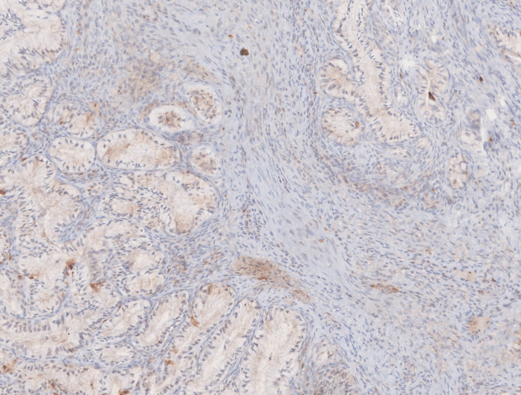
Fig.6h; p16
Immunohistochemistry for p16 was negative, done to rule out adenocarcinoma or in-situ component [Fig.6h].
Final Impression: Benign Cervical Polyp with Microglandular Adenosis.
- It is a benign, non neoplastic lesion characterized by proliferation of endocervical glands.
- Frequently associated with pregnancy & oral contraceptives intake
- May present with contact bleeding or polypoidal mass
- It simulates carcinoma due to its pseudoinfiltrative or solid pattern, presence of signet ring cells and occasional mitotic figures.
- Differential Diagnosis: Endocervical adenocarcinoma
- Immunohistochemistry:
- Positive stains: ER, PR, PAX2, Cyclin D1, p63
- Negative stains: CEA, p16, Vimentin
- Low Ki67 index
- Excellent Prognosis
Contributed by: Dr. Meenakshi Kamboj
Compiled by: Dr. Ankur Kumar & Dr. Himanshi Diwan
In case of queries, email us at: kumar.ankur@rgcirc.org
Cervix Female genital tract Microglandular adenosis Non neoplastic
Last modified: 05/06/2021