Background
A 27-year-old male presented with history of progressive shortness of breath since 2 years and noisy respiration since 1 month. CECT chest showed an ill defined heterogeneously enhancing soft tissue lesion in the pre-carinal location with small polypoidal endoluminal soft tissue component projecting into the lower trachea causing partial airway narrowing. Excision biopsy sent for examination.
Microscopy

Fig.8a; H&E; 0.65x

Fig.8b; H&E; 2.5x

Fig.8c; H&E; 5x

Fig.8d; H&E; 20x

Fig.8e; H&E; 5x

Fig.8f; H&E; 10x
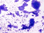
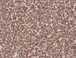
Images show a tumor in predominantly cribriform pattern lined by inner luminal and outer myoepithelial cells with punched out hyalinized or myxoid globules [Fig.8a-d]. The tumor infiltrates the tracheal cartilage [Fig.8e] and overlying respiratory mucosa [Fig.8f].
Final Impression: Adenoid Cystic Carcinoma- Grade 2.
- Salivary gland tumor composed of bilayered epithelial and myoepithelial neoplastic cells arranged in cribriform, tubular or solid pattern
- Mean age of presentation: 45 years
- Sites of involvement:
- Minor salivary glands > major salivary glands
- Most common: oral cavity
- Other sites: sinonasal tract, nasopharynx, oropharynx, trachea
- Amongst major salivary glands: Parotid is the most common site
- Most common site of metastases: Lung parenchyma
- Prognostic factors:
- Stage
- Margin status
- High mitotic index
- Solid architecture
- Genetic alteration: Fusion involving MYB, MYBL1 and NFIB genes is characteristic and diagnostic
- Treatment:
- Complete surgical resection with adequate margins with adjuvant radiotherapy.
- Chemotherapy in case of metastases
- Immunohistochemistry: Positive Stains:
- Ductal component: CK7 & CD117
- Myoepithelial component: p63, p40, Calponin, S100, SMA & CK
- Differential diagnosis:
- Epithelial myoepithelial carcinoma (cribriform pattern is focal, if present, papillocystic architecture, clear myoepithelial cells, no angulation of nucleus & HRAS mutation)
- Basal cell adenocarcinoma (shows peripheral palisading)
- Polymorphous adenocarcinoma (usually shows one type of tumor cells)
Contributed by: Dr. Meenakshi Kamboj
Compiled by: Dr. Ankur Kumar & Dr. Himanshi Diwan
In case of queries, email us at: kumar.ankur@rgcirc.org
Adenoid cystic carcinoma Head & Neck Trachea
Last modified: 05/06/2021